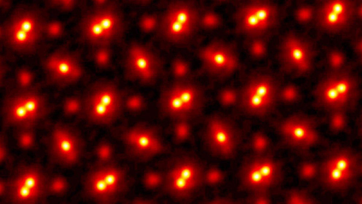
We have never seen atomic imagery in this kind of detail before
Using advanced ptychography, Cornell University researchers have captured a picoscale image 3x better than the best electron microscopy.

What you're seeing above is a praseodymium orthoscandate crystal, at 100,000,000x magnification. This is at the picometer scale, far smaller again than nanoscale that we so often think of as the bees knees of tiny tech today.
Simply put, we've never seen this kind of resolution for atomic imaging before. Not even close. The amazing team at Cornell University has actually TRIPLED the best resolution captured by any other electron microscopic system out there today.
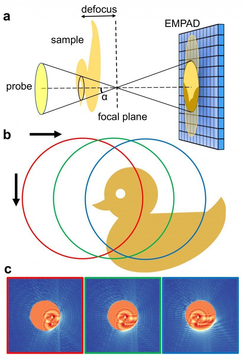
The new technique, an evolution of ptychography using an electron microscope pixel array detector (EMPAD), uses an imaging technique that is actually slightly defocused to maximise the amount of information it is receiving rather than trying to focus on one specific point. This allows for a broader set of information across three dimensions that can then be algorithmically processed to deliver this amazing picture.
The blurriness we're seeing in this picture is a result of atomic jiggle – it's just what they do.
Another massive advantage over traditional electron microscopy is that it doesn't harm the subject of the image, making these new techniques more suitable for imaging biological structures. And part of why ptychography works better is that it is capturing far more of the electrons bouncing around the system than in electron microscopy.
Take another look at those atoms. Just amazing stuff to see.
You can read a lot more about how this kind of counterintuitive system works so effectively at the Cornell University report on the research. And this rubber duck might also offer some answers.

Byteside Newsletter
Join the newsletter to receive the latest updates in your inbox.


